- HOME
- Story of the Cover
Story of the Cover
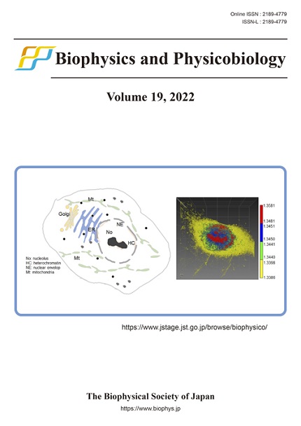
Application of quantitative cell imaging using label-free optical diffraction tomography

(Chan-Gi Pack, Biophysics and Physicobiology 18, 244-253 (2021), DOI: 10.2142/biophysico.bppb-v18.027)
The cell is spatiotemporally organized into cellular compartments, including the endoplasmic reticulum, mitochondria, nuclear envelope, and nucleolus, which have high relative molecular density. The compartments of a live HeLa cell were three-dimensionally imaged by label-free optical diffraction tomography, which allows quantification of the refractive index (i.e. molecular density) and volume of the compartments. The raw image obtained by ODT can be rendered with various refractive index (RI) ranges. The nucleolus and nuclear envelope are shown in red and the cytoplasm in yellow, respectively, with different RI ranges. The nucleoplasm is represented by two narrow RI ranges (blue and green), assuming the presence of differences in chromatin density.
 Biophysics and Physicobiology