"Visualization of peptidoglycan layer isolated from gliding diderm bacteria, Flavobacterium johnsoniae and Myxococcus xanthus, by quick-freeze deep-etch replica electron microscopy" by Yuhei O. Tahara, Tâm Mignot, Makoto Miyata is published in BPPB as the J-STAGE Advance Publication.
2025 August 26 BPPB
A following article is published as the J-STAGE Advance Publication in "Biophysics and Physicobiology".
Yuhei O. Tahara, Tâm Mignot, Makoto Miyata
"Visualization of peptidoglycan layer isolated from gliding diderm bacteria, Flavobacterium johnsoniae and Myxococcus xanthus, by quick-freeze deep-etch replica electron microscopy"
URL:https://doi.org/10.2142/biophysico.bppb-v22.0019
- Abstract
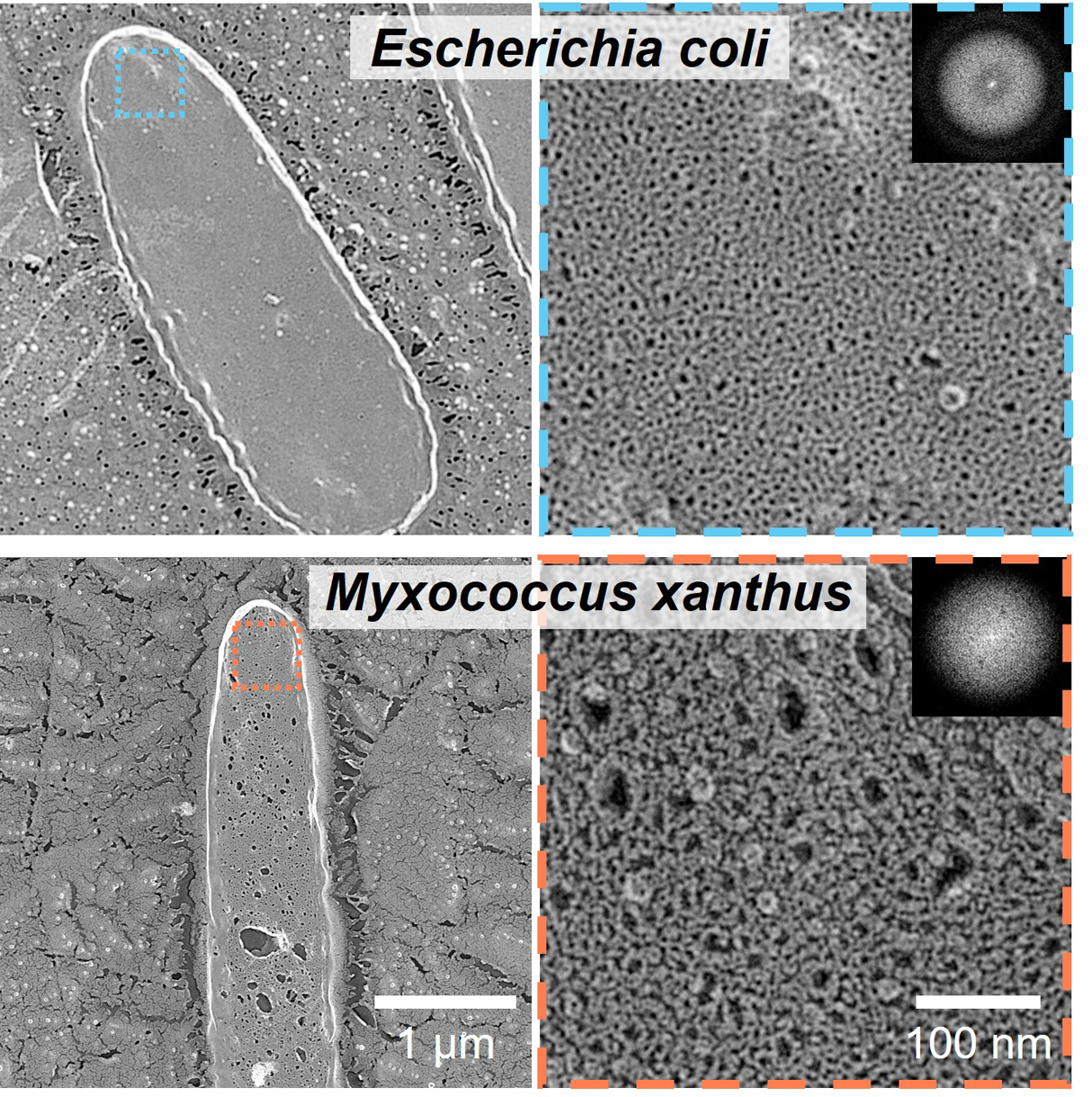
- The bacterial peptidoglycan layer plays an important role in protecting the bacteria from turgor pressure, viruses, and predators. However, it also acts as a barrier in transmitting forces generated on the cell membrane to adhesion proteins on the surface during gliding locomotion. In this study, peptidoglycan layers were isolated from two species of gliding diderm, i.e., gram-negative bacteria, and their structures were visualized by quick-freeze deep-etch replica electron microscopy. The horizontal bonding of peptidoglycan did not differ obviously among the three species. However, the diameter of pores in the peptidoglycan layer of M. xanthus and the area of surface pores were 51 nm and 14.6%, respectively, which were significantly larger than those of E. coli (32 nm and 5.8%) and F. johnsoniae (29 nm and 7.0%). Based on this, we discussed the mechanism by which diderm bacteria transmit forces across the PG layer to the bacterial surface.